Through the novel imaging technology, scientists use fluorescent imaging to locate proteins and other molecules in tissues and cells. It works by tagging the molecules with dyes that glow under specific sorts of light – the same principle behind so – known black light images.
Fluorescent imaging can assist researchers which molecules are produced in big volumes in cancer or other ailments, information that may be vital in diagnosis or in recognizing possible targets for therapeutic drugs. Looking at just one or two of the molecules in tissue or cell samples is fairly straightforward. Unfortunately, it does not offer a concise picture of how such molecules are acting in the virtual world. For this, the researchers need to expand their view.
“Biological research is moving toward intricate systems that extend across numerous dimensions, the interaction of multiple elements over time,” says postdoctoral fellow Francesco Cutrale. He introduced HySP with Scott Fraser, a USC Provost Lecturer of Biological Science who also possesses the Elizabeth Garrett Chair in Convergent Bioscience.
“By considering numerous targets, or watching targets move over time, we can obtain a much better image of what is actually happening within intricate living system,” says Cutrale. Presently, scientists must consider various labels separately, and then apply intricate methods to layer them together and identify how they related to each other, a time-consuming and costly procedure, Cutrale says. HySP can consider numerous distinct molecules in single pass.
“Consider looking at 18 targets.” says Cutrale. “We can perform them all at once, rather than having to perform 18 separate studies and try to link them later.” Moreover, the algorithm efficiently filters through interference to discern the real signal, even if that signal is entirely weak, very much like identifying the proverbial needle in a haystack. Present technology from NASA’s Jet Propulsion Lab can also perform this, but the process and equipment are both extremely costly and time-consuming.
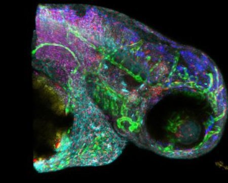
“HySP employs much less computing time, and we do not require the costly imaging instrumentation,” says Fraser. Fraser and Cutrale, along with the scientists from Keck School of Medicine of USC, Caltech, and the University of Cambridge in UK have employed zebra fish to identify and introduce HySP. In this common lab model, the system works entirely well. But the concern is about the people.
Conclusion
The researchers expect to test the process in the next couple of years with the aid of soldiers whose lungs have been damaged by chemicals and irritants they might have experienced in combat. The scientists will extend a light releasing probe into the lungs of soldiers that will record images of the fluorescence in the encompassing tissues. They will they employ HySP to prepare what volumes to a fluorescent map and compare it with that of healthy lung tissue to identify if they can discern the damage.
Filed Under: News

Questions related to this article?
👉Ask and discuss on EDAboard.com and Electro-Tech-Online.com forums.
Tell Us What You Think!!
You must be logged in to post a comment.