One of the few compulsory gadgets that every doctor carries is the Stethoscope. A sophisticated assembly constructed out of steel and plastic, stethoscope is an age-old source to diagnose cardiac problems. Popularly known as “Doctor’s Weapon”, stethoscopes have seen innumerous changes in their structure and working since their invention in 1824. Models that can be tuned with a smart phone (yes, really!) have already hit the market. Peculiarly, almost every type of stethoscope has found its space in the vast world of medical experts. There’s one for a cardiac for example, a different one for a pediatrician and yet another for a physicist.
This Insight covers one of the most popular types of stethoscopes: Littmann Stethoscopes, which you may recognize as the serpentine devices hanging around doctors in movies. These stethoscopes are one of the oldest species and are still recommended for their precise results.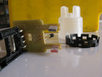
Fig. 1: Image Showing A Typical Littmann Stethoscope
Stethoscope Assembly and Headset
Image 02 shows a conventional acoustic Littmann stethoscope. This stethoscope is a simple assembly of three: a chest piece, hollow rubber tube and a steel frame. The assembly must be handled with extreme care: application of a slightly more cleaning agent on its surface can give erroneous results.
Fig. 2: Various Parts of A Stethoscope
Image 03 details the headset region of the stethoscope. The steel tubes, also called as ear-tubes, are pliable so that headsets can easily be worn by all. Also, once the ear knobs are plugged to the ears, the headset region resides in the front without being required to be held by the user. The ear knobs get placed right at the opening of ear canal making the user get maximum output.
Ear Knobs

Fig. 3: Closer View of Rubber Ear Knobs in A Stethoscope
Ear knobs (a.k.a ear pieces) are made of soft rubber, thus providing a comfortable cushion comfort to the ears. Once, placed in the ear canal, they seal it off, thus preventing any outside noises to disturb the observer.
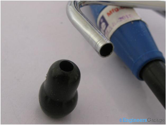
Fig. 4: Image Showing The Edge of Hollow Ear Tube Under Ear Knobs
The ear knobs can be taken off to reveal the hollow structure of the steel eartubes. The eartubes are ribbed or well furnished so that they can resist mechanical abrasions to an extent.
Steel Eartubes and Frame

Fig. 5: Image Showing Extended Steel Tube Under Rubber Tube
The steel eartubes extend to the rubber tubes which can be incised to see the extent to which the steel eartubes are placed in the stethoscope.
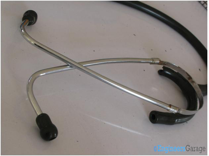
Fig. 6: Ear Tubes Steel Frame
Incisions on the rubber tube make it easy to remove the steel eartubes that extend to form a frame.

Fig. 7: Image Showing Patterns of Steel Frame
Image 08 shows the manner in which the eartubes extend to form the frame. The manner in which the circular tube extends to form a flat surface that forms a steel frame structure for the headset is distinctly shown in the picture.
The flat region of the frame gives the flexible property to the frame and also holds the eartubes firmly when in use.
Acoustic Rubber Tube

Fig. 8: A Closer Look At Rubber Tube and Chest Piece
Except a few incision marks on the rubber tube, there are no significant damages to the chest piece and rubber tube. Let’s find out the intricacies of these two crucial parts of the stethoscope.

Fig. 9: Detailed Look Of Rubber Tube
The rubber tube is thick (double lumen) structure which is flexible enough to get bent at any angle without causing any mechanical damage. The task of this tube is to convey the acoustics generated by the chest piece to the headset. It is glued permanently to the chest piece and the headset, the only manner to detach them off is through incisions.

Fig. 10: Side View of Stethoscope Chest Piece
Side view of the chest piece of the stethoscope is seen in this mage. Light weighted, so that it is comfortable to carry, the chest piece is made from Aluminum whose outer coat is anodized.
Bell & Diaphragm Region of Chest Piece
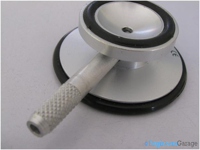
Fig. 11: The Sensing Part of Stethoscope’s Chest Piece

Fig. 12: Diaphragm or Sensing Part of Chest Piece
Images 012 and 013 show the two sensing regions of the chest piece: the bell and the diaphragm. Stethoscope can be made to work with one of them at a time by rotating a rod attached to piece. The rod works as a knob and tunes the stethoscope to the sensing region where it is desired to work.
The stethoscope is tuned to the diaphragm region when high frequency sounds (auscultation of adult patients) are to be sensed and it is tuned to the bell region in the cases of low frequency sounds (auscultation of young or extremely thin patients).
Both the sides have a plastic ring at their circumferences so that the user does not feel the chill of the metallic region of the chest piece.
Diaphragm and Chest Piece Screws

Fig. 13: Various Parts of Stethoscope’s Diaphragm
The diaphragm is a thin plastic sheet, designed to detect even slightest of the acoustic changes that it senses in its vicinity. The acoustic changes occur in form of pressure on the surface of diaphragm.
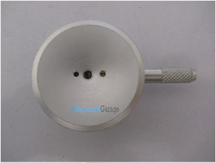
Fig. 14: Stethoscope’s Bare Chest Piece
After removal of the diaphragm and the rubber ring around it, bare chest piece is seen. The diaphragm and the bell have a small hole each from where the acoustic signals reach the earpiece. In addition to that, the diaphragm region has one-two additional small holes. These additional ones aid in tightening the knob to the chest piece and enable easy twisting.
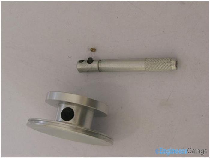
Fig. 15: Disassembled Chest Piece of Stethoscope
The screw has been taken out to detach the tightening knob and the chest piece. Usually, the knob and the chest piece are considered a single entity.
Filed Under: Insight

Questions related to this article?
👉Ask and discuss on Electro-Tech-Online.com and EDAboard.com forums.
Tell Us What You Think!!
You must be logged in to post a comment.